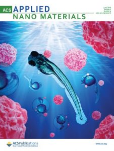
Featured paper: Low toxicity magneto-luminescent nanoplatform.

Cover of ACS Applied Nano Materials features the paper.

In research carried out in a number of Brazilian laboratories, a multidisciplinary scientific team developed a magnetic, luminescent nanomaterial capable of chemically binding to molecules of interest, such as drugs or proteins. This nanomaterial also showed low toxicity in tests with living organisms. With this set of characteristics, the new material can be seen as a multifunctional nanoplatform that is promising for the development of various applications, especially in the areas of biotechnology, health and environment. The study was reported in an article published in ACS Applied Nano Materials (American Chemical Society journal released in 2018), and featured on the cover of the June issue of the journal.

The properties of this nanoplatform derive from the presence of several compounds and elements with distinct properties: iron oxide (Fe3O4, known as magnetite) nanoparticles responsible for magnetism; lanthanide element ions (Gd3 +, Ce3 + and Tb3 +, known as rare earths) responsible for luminescence or light emission, and chitosan (biopolymer obtained from the crustacean exoskeleton), essential for providing chemical bonds of the nanoplatform surface to the external molecules of interest.

The nanoplatform was developed at the Brazilian National Nanotechnology Laboratory of the National Center for Energy and Materials Research (LNNano – CNPEM). The process used for its synthesis comprises a series of steps. Initially, the iron oxide nanoparticles that form the core of the nanoplatforms are synthesized and coated with silicon dioxide (SiO2). Then the luminescent elements and chitosan are incorporated into the nanoparticles forming an outer layer. The result is nanoplatforms of approximately 170 nm in diameter (on average), called Fe3O4@SiO2/GdOF:xCe3+,yTb3+.
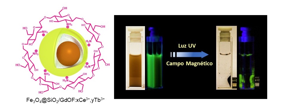
On the left, a schematic illustration of one of the nanoplatforms, showing its core. On the right, a solution with nanoplatforms under the effect of a magnetic field (concentrated near the magnets) and irradiated with UV light (generating the emission of green light).

To study the magnetic and luminescent properties of the nanoplatform and to characterize its structure and morphology, research groups from the State University of Campinas (Unicamp) and the University of São Paulo (USP) participated in the study.

In addition, the main authors of the paper decided to evaluate the toxicity of nanoplatforms with relation to living organisms – a key step when thinking about health or environmental applications, and they decided to conduct a well-established in vivo test, in which zebrafish embryos (scientific name Danio rerio) are exposed to the material whose toxicity is to be evaluated. These freshwater fish, in fact, has a high genetic similarity to humans (about 70%) and at the same time is cheaper and easier to study than mice or rats, among other advantages.

In the toxicity test, a few dozen freshly fertilized zebrafish eggs were placed in aqueous medium containing the nanoplatforms at various concentrations. The embryos were examined at different development stages using an optical microscope to check for mortality, malformation, edema or changes in size. Tests included embryos with and without chorion (membrane that protects the embryo in the early stages of development). The test results carried out at LNNano showed that nanoplatforms, even at high concentrations (100 mg/L), have low toxicity for all embryo groups.
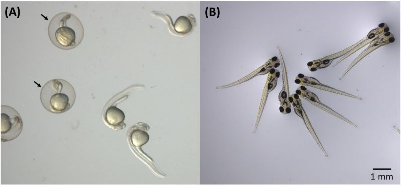
Zebrafish embryos used in nanotoxicity tests. (A) 24-hour embryos in the presence and absence of the chorion, where arrows indicate the chorion (membrane that protects embryos in the early stages of development). (B) Embryos after 96 hours of development.

“This work brings an unprecedented contribution that involves evaluating the toxicity of hybrid nanomaterials using the zebrafish model, a promising alternative method in nanotoxicology, and the influence of the chorion,” says Diego Stéfani Teodoro Martinez, CNPEM researcher at LNNano and one of the corresponding authors of the article.

The embryos were also analyzed at the Brazilian National Synchrotron Light Laboratory (LNLS – CNPEM) to verify the distribution and concentration of nanoplatforms in the organism of the embryos. To do this, the scientists used the synchrotron light X-ray fluorescence microscopy (SXRF) technique, which can accurately map certain chemical elements in biological systems. This technique is available at one of the LNLS experimental stations, coordinated by the researcher Carlos Alberto Pérez, who is one of the corresponding authors of the article.
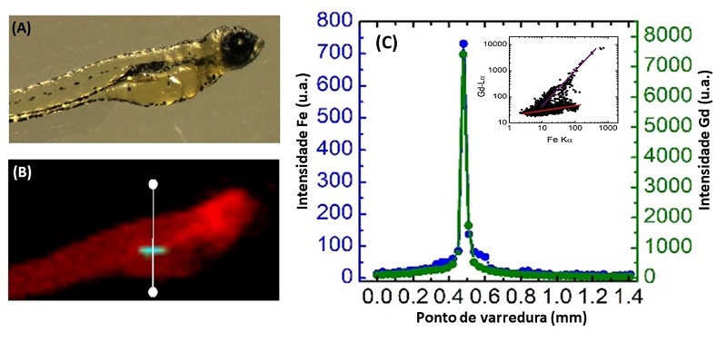
X-ray fluorescence microscopy analysis with synchrotron light (SXRF) of two zebrafish embryos after exposure to the nanoplatform for 72 hours. (A) Optical microscopy image of the embryos; (B) SXRF image of embryos demonstrating nanoplatform accumulation in the intestinal tract; and (C) X-ray fluorescence intensity along the white line indicated in (B), demonstrating the spatial co-location of Fe and Gd elements in the intestinal tract of zebrafish embryos.

SXRF analysis showed that nanoplatforms had accumulated in the embryos as a function of exposure time, with higher concentrations in the gastrointestinal tract in the case of already developed mouth embryos – a result that may be significant, for example in the context of healthcare applications involving oral nanoplatform ingestion.

The study was carried out in the context of a postdoctoral project by fellow Latif Ullah Khan, also corresponding author of the article. The completion of the project, says Martinez, was made possible by the availability of skills and facilities at CNPEM’s multi-user laboratories. However, partnerships with other laboratories were also crucial, adds the CNPEM researcher. Professor Marcelo Knobel’s group performed the magnetometry studies at Unicamp. The groups of professors Hermi Felinto Brito and Magnus Gidlund carried out the luminescence and functionalization studies at USP. Finally, Professor Diego Muraca (Unicamp) and researcher Jefferson Bettini (CNPEM) contributed to the structural and morphological characterization using transmission electron microscopy techniques.

“This article was the result of integrating the experience of different Brazilian groups; an interdisciplinary study on the frontier of knowledge in nanobiotechnology and nanotoxicology,” says Martinez, adding that one of the main challenges of the work was integrating knowledge and techniques from different areas, such as Materials, Biology and Toxicology, a task that was coordinated by Martinez and Pérez.
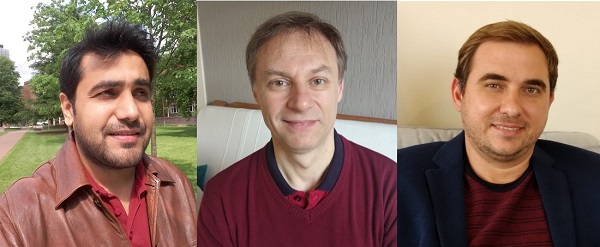
The main authors of the paper. From the left: Latif Khan, Carlos Pérez and Diego Stéfani Martinez.

The study received financial support from Brazilian agencies CAPES (including through the CAPES-CNPEM agreement), FAPESP and CNPq (including through INCT-Inomat); from the Brazilian Ministry of Science, Technology, Innovations and Communications (MCTIC) through SisNANO, and The World Academy of Sciences for advancement of science in developing countries (TWAS). The study also received financial support from the Brazil-China Nanotechnology Research and Innovation Center (CBC-Nano).

Applications: biotechnology, health and the environment

According to Martínez, the nanoplatform developed opens perspectives for applications in biotechnology, health and the environment, such as biological tissue and cell imaging systems, medical diagnostic kits, and environmental systems for pollutant detection and remediation

The applications would take advantage of the interesting set of nanoplatform properties. Because they are magnetic, using an external magnet, nanoplatforms could be directed and retained in a particular biological tissue or isolated from, for example, contaminated blood or water. In addition, the luminescence of the nanomaterial would allow visualizing the nanoplatforms within the biological tissues and cells of interest. Finally, the presence of chitosan would enable the chemical binding of drugs and other molecules that would serve for the diagnosis and/or treatment of diseases. “However, much study is still needed for real applications and commercialization of this nanoplatform, as it is a new material and needs to be tested on different models in the future,” says Martinez Martinez.


[Paper: Fe3O4@SiO2 Nanoparticles Concurrently Coated with Chitosan and GdOF:Ce3+,Tb3+ Luminophore for Bioimaging: Toxicity Evaluation in the Zebrafish Model. Latif U. Khan, Gabriela H. da Silva, Aline M. Z. de Medeiros, Zahid U. Khan, Magnus Gidlund, Hermi F. Brito, Oscar Moscoso-Londoño, Diego Muraca, Marcelo Knobel, Carlos A. Pérez, Diego Stéfani T. Martinez. ACS Appl. Nano Mater. 2019, 2,6, 3414-3425. https://doi.org/10.1021/acsanm.9b00339.]

SBPMat newsletter. English edition. Year 3, issue 6.


Brazilian Materials Research Society (SBPMat) newsletter
News update from Brazil for the Materials community

English edition. Year 3, issue 6. 

XV Brazil-MRS (SBPMat) Meeting - Campinas (SP), Sept 25-29, 2016 

The XV SBPMat Meeting received approximately 2,000 abstracts.

Registration: Registration for the event is now open. Early registration discount deadline is 31 August. Here.

Program: Two tutorials will be offered on the afternoon of September 25 to those registered for the event at no extra cost. One is on computer simulations on atomic systems using Reactive Force Fields (theory and practice). The second, organized by Professor Valtencir Zucolotto, will address capabilities required to make high-impact science, including scientific writing. Reserve your place during registration. 

Authors: Acceptance notifications will be sent to the authors by July 10. 

Awards: Those interested in participating in the event’s student prize competition, the Bernhard Gross Award, which selects one oral and one poster presentation in each symposium, must submit an extended abstract by August 22. Know more in the instructions to authors.  

Publication of contributions: The papers presented at the XV Brazil-MRS Meeting may be submitted by their authors for peer review for publication in IOP scientific journals. More info.

Exhibitors: More than 30 companies have already got places in our exhibition. Companies interested in participating in the event with stands and other forms of dissemination should contact Alexandre, via the e-mail comercial@sbpmat.org.br.

Plenary sessions:  View the abstracts of the plenary lectures and the memorial lecture of our event and bios of the scientists presenting them. Here.

Accommodation and tickets: See the list of the travel agency “Follow Up” with hotels, hostels, guesthouses and the forms to book flights. Here. 

Vacation packages: The Follow Up website also suggests tour packages for before and after the event. Here.

Venue: See video of the city of Campinas and folder about the Expo Dom Pedro convention center. 

Organizers: This edition of the event is coordinated by Prof. Ana Flávia Nogueira (Unicamp, Institute of Chemistry) and Prof. Mônica Alonso Cotta (Unicamp, “Gleb Wataghin” Institute of Physics). See who are the members of the local committee and view the photos of the organizers. Here.

Featured paper 

A nanomedicine study performed at the Brazilian Federal University of Goias shows that magnetic nanoparticles smaller than 10 nm and composed of more than one material have optimum nano-heating properties for the treatment of cancer by hyperthermia. The two authors of the study reached these conclusions based on diverse evidence, including in vivo studies and results obtained through an innovative theoretical method that they developed. This work was reported in a paper published in Nanoscale. See our story about the study.

People in the Materials community 

We interviewed professor Sidney Ribeiro (UNESP), a full member of the Brazilian Academy of Sciences since May. Ribeiro is a recognized author of impacting studies on materials containing rare earth ions with applications in photonics and biomedicine. He is also active in mentoring and training researchers (having supervised over one hundred studies) and transforming research into products. In his message to younger scientists he spoke about the love of science, which is natural in children and must be preserved by the educational system, and which transforms the researcher’s work into a favorite occupation. See our interview.

Professor Fernando Lázaro Freire Junior, former president of SBPMat, became the director of the Physics Department of PUC-Rio. Here.
Interviews with plenary speakers of the XV Brazil-MRS Meeting
Plants and animals are important sources of knowledge and inspiration for Professor Lei Jiang and his group. In their laboratories at the Technical Institute of Physics and Chemistry in Beijing (China), they develop smart materials, e.g., interfaces that switch between superhydrophilicity and superhydrophobicity. The findings of professor Lei Jiang, in addition to generating publications that received tens of thousands of citations, yielded products which are already widely used. Learn more about this Chinese scientist, his way of doing science, his discoveries and his scientific and also philosophical concept of binary cooperative complementary materials. Here.
Special: Kavli Prize for AFM inventors
Gerd Binnig (IBM Zurich Research Laboratory, Switzerland), Christoph Gerber (University of Basel, Switzerland) and Calvin Quate (Stanford University, USA) received the 2016 Kavli Prize in Nanoscience in recognition of their development of the Atomic Force Microscope (AFM). Since its creation, the AFM advances nanoscience and nanotechnology due to the possibilities it offers to study and modify surfaces with atomic resolution and precision. More. 
Reading tips
  • First stable magnet of only 1 atom provides possibilities to store and process information at the atomic scale (based on paper in Science). Here.
  • Biomineralization: Scientists shed light on the origin of hardness in biominerals as calcite, associated to the incorporation of impurities (based on paper in Nature Materials). Here. 
  • Thomson Reuters released its annual report of scientific journal impact factors. Here are some highlights of materials journals selected by the websites Materials Today (Elsevier) and Materials Views (Wiley)
Events
  • XXV International Conference on Raman Spectroscopy (ICORS2016). Fortaleza, CE (Brazil). August, 14 to 19, 2016. Site.
  • 26th LNLS Annual Users´ Meeting (RAU). Campinas, SP (Brazil). August, 24 to 25, 2016. Site.
  • XV Brazil-MRS Meeting (XV Encontro da SBPMat). Campinas, SP (Brazil). September, 25 to 29, 2016. Site.
  • Aerospace Technology 2016. Stockholm (Sweden). October, 11 to 12, 2016. Site.


To unsubscribe, click here


Featured paper: Best nanoheaters for cancer treatment.

[Paper: Mean-field and linear regime approach to magnetic hyperthermia of core-shell nanoparticles: can tiny nanostructures fight cancer? Marcus S. Carrião, Andris F. Bakuzis. Nanoscale, 2016,8, 8363-8377. DOI: 10.1039/C5NR09093H]

The authors of the paper published in Nanoscale: Professor Andris Bakuzis (left) and doctoral student Marcus Carrião dos Santos (right).

Hyperthermia, for cancer treatment, is induced by increasing the temperature to activate tumor cell death. This high temperature can be created by introducing nanoparticles into the tumor that function as heaters, and after their function is completed they are eliminated by the body. Magnetic nanoparticles may be used in these treatments because they have the ability to generate heat when subjected to alternating magnetic fields of specific intensity and frequency.

A work on nanomedicine (nanotechnology used in medicine) fully conducted at the Brazilian Federal University of Goiás (UFG) suggests a new strategy using hyperthermia for cancer treatment: using smaller magnetic nanoparticles than those normally used and composed of more than one material, which could provide several advantages to the patient. To reach this conclusion, the researchers developed an innovative theoretical method that paves the way for manufacturing magnetic nanoparticles optimized for hyperthermia. The study was reported in a paper published in the prestigious Nanoscale journal, signed by the doctoral student Marcus Carrião dos Santos and his supervisor Andris Figueiroa Bakuzis, professor at the Physics Institute of UFG.

Hyperthermia cancer treatment generally uses nanoparticles that are relatively large (20 nm size range) and homogeneous (from a single material), which are considered as the most effective to generate heat according to theoretical studies based on traditional methods. However, these “large” nanoparticles accumulate quickly in the liver and it may take several months or years for these particles to leave the body of the patient being treated. On the other hand, nanoparticles smaller than 10 nm are rapidly eliminated in the urine, reducing the possibilities of intoxication and thereby increasing the selection of materials that can be used to manufacture them.

The relationship between particle size and excretion route (liver or kidney) was a conclusion reached by Bakuzis and colleagues from evidence reported in the scientific literature and pre-clinical studies (in vivo) carried out within a multidisciplinary research network, coordinated by Bakuzis, and aimed at solving problems associated with the use of magnetic nanoparticles for cancer treatment.

In addition, smaller nanoparticles have better distribution and penetration in tumors, among other advantages in the context of cancer treatment.

Aware of these characteristics, Bakuzis and dos Santos investigated the possibility of manufacturing nanoparticles of less than 10 nm that could efficiently generate heat. An important insight came from an article published in 2011 in the Nature Nanotechnology journal (Nat. Nanotech. 6, 418 (2011)). Professor Bakuzis says that “in this article, the researchers concluded experimentally that certain heterogeneous (from different materials) core-shell structures of spinel ferrites warmed more efficiently than homogeneous particles”.

The scheme, provided by the authors, sums up the hyperthermia process of magnetic nanoparticles and compares the conventional nanoparticles with the proposal of the UFG researchers, showing the key advantages for its application in the treatment of cancer using hyperthermia.

The pair of scientists then decided to theoretically investigate whether nanoparticles less than 10 nm formed by one material core and a shell of another material could efficiently generate heat and how to optimize them for this function. However, the conventional methods available for this modeling were not adequate. In fact, they considered the nanoparticle as a homogeneous entity, ignoring the fact that the surface atoms and the core atoms respond differently to the application of a magnetic field. This oversight became more significant in the study of particularly heterogeneous particles such as those they intended to study, the reason why the researchers from Goiás decided to develop a more suitable model for the study object.  Bakuzis explains that “in the paper we presented the first analytical hyperthermia model of core-shell nanoparticles within the linear response and mean-field theory, and from these calculations we pointed out important materials properties to achieve efficient heat generation.”

The results obtained by the physicists and published in the paper may have a significant impact in a health issue that concerns humanity, cancer cure. “Our studies indicate that it is possible to develop small particles for cancer treatment that can be quickly eliminated from the body via the kidneys. In particular, by combining different materials in the nanostructure”, summarizes Bakuzis.

To work with impact on this interface theme, Bakuzis is always in contact with a pool of knowledge of various areas. In addition to leading the multidisciplinary nanomedicine network that includes researchers with backgrounds in tumor biology, genetics, physiology, pharmacy, veterinary medicine, biophysics, physics, medical physics and chemistry, the professor and his group actively participate in scientific events that bring together many different professionals, including doctors with various specializations already using hyperthermia in humans for cancer treatment. “These scientific contacts are fundamental in interface areas such as the one our group works with,” concludes Bakuzis.

The research that led to the paper in the Nanoscale journal received funding from the Brazilian National Scientific and Technological Development Council (CNPq) and from the Research Foundation of the State of Goiás (FAPEG) and was carried out as part of the doctoral work of Marcus Carrião dos Santos.