Featured paper: A molecular machine to fight cancer.

[A reversible, switchable pH-driven quaternary ammonium pillar[5]arene nanogate for mesoporous silica nanoparticles. Santos, ECS ; dos Santos, TC; Fernandes, TS; Jorge, FL; Nascimento, V; Madriaga, VGC ; Cordeiro, PS; Checca, NR; Da Costa, NM; Pinto, LFR; Celia Ronconi. J. Mater. Chem. B, 2020,8, 703-714. https://doi.org/10.1039/C9TB00946A]

A molecular machine to fight cancer

In 2016, the smallest man-made machines ever created, called molecular or nanomachines, gained visibility with the Nobel Prize in Chemistry. These nanometer-sized machines, whose components are molecules that perform controlled movements, could help humanity accomplish complex tasks at the molecular scale.

In the health area, one such task is to effectively fight cancer cells without damaging healthy tissues. It is known that nowadays one of the main problems of the most used therapies concerns the side effects on healthy tissues – a problem that has led many scientists to develop drug delivery systems that can take drugs directly to cancer cells without leaking.

At the Brazilian Federal Fluminense University (UFF), over the last ten years Professor Célia Machado Ronconi and her scientific team have been working on nanomachines for cancer treatment. In her postdoctoral research, carried out between 2003 and 2005, the scientist learned about molecular machines at the University of California, Los Angeles (UCLA), at one of the most qualified laboratories in the world working on this subject – the research group of Sir James Fraser Stoddart, who years later would be awarded the Nobel Prize mentioned at the beginning of this article, alongside with Jean-Pierre Sauvage and Bernard L. Feringa.

In a recently published paper in Journal of Materials Chemistry B, Professor Célia Ronconi, her team and collaborators, all from Brazilian institutions, presented a new nanomachine composed of a drug reservoir and a cap. The machine has an opening/closing lid mechanism that responds to changes in the acidity of the medium in which it is located. When the pH of the medium is similar to that of the blood of a healthy human being (physiological medium), the cap remains closed, preventing the drug from being released. When the pH is more acidic, a characteristic seen around cancer cells, the lid opens and the drug is released. In laboratory in vitro tests, the nanomachine loaded with a well-known chemotherapeutic drug proved to be more effective than the pure drug in eliminating breast cancer cells, destroying 92% of them in 48 hours.

The highlight of this figure shows a zoom of the nanomachine loaded with the drug (green balls). The zoom focuses one of the nanochannels of the closed reservoir and its nanocap, preventing the drug from being released.

With these characteristics, the nanomachine developed at UFF shows application potential in the delivery of chemotherapeutic drugs to cancer cells. “The results of this work were extremely promising,” says Professor Ronconi. “However, there is still much to be studied. The next steps of the work will be to test the nanomachine loaded with the drug in other breast cancer cell lines, as only one line (MCF-7) was tested. We will also test the toxicity of the device without the drug in healthy cells and, if the results are positive, in vivo studies will be carried out, using mices genetically altered to have a deficient immune system, ” adds Professor Ronconi.

Assembly and operation of the nanomachine

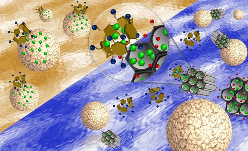
To achieve the reservoir function, the UFF group synthesized spherical mesoporous silica nanoparticles of about 85 nm in diameter. In addition to being biocompatible, this material has a unique internal honeycomb-like structure, with a set of nanochannels of up to 4 nm in diameter, in which the drug molecules can be stored. The nanoparticles were covered with carboxyl groups (- COOH) that improved the interaction of the reservoir with its cap. For the cap, the researchers chose pilararene, an artificial molecule made up of five aromatic rings, whose first synthesis dates back to 2008 in the scientific literature.

In the assembly and operation of the nanomachine, the electrostatic interactions of attraction controlled by the medium pH were the great allies of the scientific team at UFF. In fact, as confirmed by the researchers in their experiments, in a solution with a pH of 7.4, which represents the acidity of healthy blood, the carboxyl groups (-COOH) that cover the reservoir lose a proton forming carboxylate groups (-COO- ), negatively charged, which interact electrostatically with the positively charged cap. Thus, the electrostatic attraction brings the two parts of the nanomachine together until it prevents the drug from being released. By lowering the pH, that is, by making the solution more acidic, the carboxylate groups (-COO-) gain protons, neutralizing their charge. As a result, the electrostatic attraction between the cap and the reservoir breaks apart, the cap opens and the drug is released.

Functioning of the nanomachine loaded with the drug (pink balls). On the left, at physiological pH, the lids close the reservoir's nanochannels. On the right, the more acidic medium generates the removal of the caps and the drug is released.
Functioning of the nanomachine loaded with the drug (pink balls). On the left, at physiological pH, the lids close the reservoir’s nanochannels. On the right, the more acidic medium generates the removal of the caps and the drug is released.

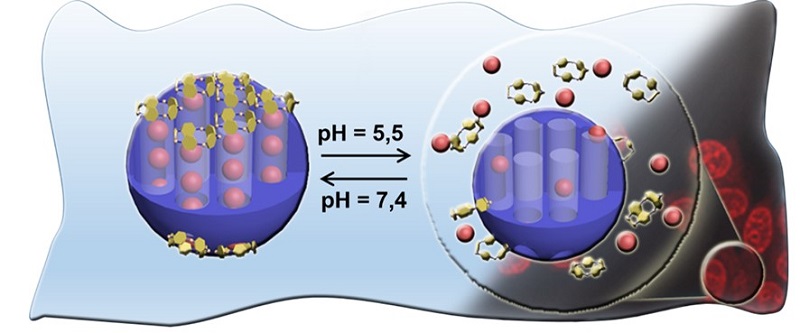
In the experiments carried out, the UFF group was able to partially release the chemotherapeutic drug (34%) at a pH of 5.5 (probably similar to that surrounding the cancer cells) and almost totally (91%) in a 2.0 acidity medium. All experiments were carried out at a temperature of 37 °C, similar to that of the human body.

History of work

Since 2009, when she became a professor at UFF and set up the Laboratory of Supramolecular Chemistry and Nanotechnology, Professor Célia Ronconi has been working in the different development phases of diverse nanomachines and drug transport and release systems, using chemical, magnetic and luminous stimulants. During Evelyn da Silva Santos’ doctorate, under the guidance of Ronconi, a nanomachine prototype was developed using material available on the market. However, new studies carried out after the defense of her doctorate work, in 2018, showed that the nanoparticles used as reservoirs formed clusters in the physiological environment (the solution that emulates blood in experiments). Thus, Professor Ronconi involved postdoctoral fellow Thiago Custódio dos Santos and doctoral student Tamires Soares Fernandes in the development of new material. “They continued the project and synthesized a material with excellent dispersion in the physiological environment, and the device was redone, as well as the drug release studies,” says professor Ronconi. The biological tests of the nanomachine were performed at INCA’s molecular carcinogenesis group, by researchers Luis Felipe Ribeiro Pinto and Nathália Meireles da Costa, and the technician Fernanda Jorge. The study also included the participation of the Brazilian Center for Research in Physics (CBPF) in the characterization of materials by microscopy techniques, carried out at the Multi-User Laboratory for Nanoscience and Nanotechnology (LABNANO). The research received funding from the Brazilian agencies CNPq, CAPES and FAPERJ.

Main authors. From the left: Evelyn Santos, Thiago Custódio, Tamires Soares and Célia M. Ronconi.

SBPMat newsletter. English edition. Year 3, issue 6.


Brazilian Materials Research Society (SBPMat) newsletter
News update from Brazil for the Materials community

English edition. Year 3, issue 6. 

XV Brazil-MRS (SBPMat) Meeting - Campinas (SP), Sept 25-29, 2016 

The XV SBPMat Meeting received approximately 2,000 abstracts.

Registration: Registration for the event is now open. Early registration discount deadline is 31 August. Here.

Program: Two tutorials will be offered on the afternoon of September 25 to those registered for the event at no extra cost. One is on computer simulations on atomic systems using Reactive Force Fields (theory and practice). The second, organized by Professor Valtencir Zucolotto, will address capabilities required to make high-impact science, including scientific writing. Reserve your place during registration. 

Authors: Acceptance notifications will be sent to the authors by July 10. 

Awards: Those interested in participating in the event’s student prize competition, the Bernhard Gross Award, which selects one oral and one poster presentation in each symposium, must submit an extended abstract by August 22. Know more in the instructions to authors.  

Publication of contributions: The papers presented at the XV Brazil-MRS Meeting may be submitted by their authors for peer review for publication in IOP scientific journals. More info.

Exhibitors: More than 30 companies have already got places in our exhibition. Companies interested in participating in the event with stands and other forms of dissemination should contact Alexandre, via the e-mail comercial@sbpmat.org.br.

Plenary sessions:  View the abstracts of the plenary lectures and the memorial lecture of our event and bios of the scientists presenting them. Here.

Accommodation and tickets: See the list of the travel agency “Follow Up” with hotels, hostels, guesthouses and the forms to book flights. Here. 

Vacation packages: The Follow Up website also suggests tour packages for before and after the event. Here.

Venue: See video of the city of Campinas and folder about the Expo Dom Pedro convention center. 

Organizers: This edition of the event is coordinated by Prof. Ana Flávia Nogueira (Unicamp, Institute of Chemistry) and Prof. Mônica Alonso Cotta (Unicamp, “Gleb Wataghin” Institute of Physics). See who are the members of the local committee and view the photos of the organizers. Here.

Featured paper 

A nanomedicine study performed at the Brazilian Federal University of Goias shows that magnetic nanoparticles smaller than 10 nm and composed of more than one material have optimum nano-heating properties for the treatment of cancer by hyperthermia. The two authors of the study reached these conclusions based on diverse evidence, including in vivo studies and results obtained through an innovative theoretical method that they developed. This work was reported in a paper published in Nanoscale. See our story about the study.

People in the Materials community 

We interviewed professor Sidney Ribeiro (UNESP), a full member of the Brazilian Academy of Sciences since May. Ribeiro is a recognized author of impacting studies on materials containing rare earth ions with applications in photonics and biomedicine. He is also active in mentoring and training researchers (having supervised over one hundred studies) and transforming research into products. In his message to younger scientists he spoke about the love of science, which is natural in children and must be preserved by the educational system, and which transforms the researcher’s work into a favorite occupation. See our interview.

Professor Fernando Lázaro Freire Junior, former president of SBPMat, became the director of the Physics Department of PUC-Rio. Here.
Interviews with plenary speakers of the XV Brazil-MRS Meeting
Plants and animals are important sources of knowledge and inspiration for Professor Lei Jiang and his group. In their laboratories at the Technical Institute of Physics and Chemistry in Beijing (China), they develop smart materials, e.g., interfaces that switch between superhydrophilicity and superhydrophobicity. The findings of professor Lei Jiang, in addition to generating publications that received tens of thousands of citations, yielded products which are already widely used. Learn more about this Chinese scientist, his way of doing science, his discoveries and his scientific and also philosophical concept of binary cooperative complementary materials. Here.
Special: Kavli Prize for AFM inventors
Gerd Binnig (IBM Zurich Research Laboratory, Switzerland), Christoph Gerber (University of Basel, Switzerland) and Calvin Quate (Stanford University, USA) received the 2016 Kavli Prize in Nanoscience in recognition of their development of the Atomic Force Microscope (AFM). Since its creation, the AFM advances nanoscience and nanotechnology due to the possibilities it offers to study and modify surfaces with atomic resolution and precision. More. 
Reading tips
  • First stable magnet of only 1 atom provides possibilities to store and process information at the atomic scale (based on paper in Science). Here.
  • Biomineralization: Scientists shed light on the origin of hardness in biominerals as calcite, associated to the incorporation of impurities (based on paper in Nature Materials). Here. 
  • Thomson Reuters released its annual report of scientific journal impact factors. Here are some highlights of materials journals selected by the websites Materials Today (Elsevier) and Materials Views (Wiley)
Events
  • XXV International Conference on Raman Spectroscopy (ICORS2016). Fortaleza, CE (Brazil). August, 14 to 19, 2016. Site.
  • 26th LNLS Annual Users´ Meeting (RAU). Campinas, SP (Brazil). August, 24 to 25, 2016. Site.
  • XV Brazil-MRS Meeting (XV Encontro da SBPMat). Campinas, SP (Brazil). September, 25 to 29, 2016. Site.
  • Aerospace Technology 2016. Stockholm (Sweden). October, 11 to 12, 2016. Site.


To unsubscribe, click here


Featured paper: Best nanoheaters for cancer treatment.

[Paper: Mean-field and linear regime approach to magnetic hyperthermia of core-shell nanoparticles: can tiny nanostructures fight cancer? Marcus S. Carrião, Andris F. Bakuzis. Nanoscale, 2016,8, 8363-8377. DOI: 10.1039/C5NR09093H]

The authors of the paper published in Nanoscale: Professor Andris Bakuzis (left) and doctoral student Marcus Carrião dos Santos (right).

Hyperthermia, for cancer treatment, is induced by increasing the temperature to activate tumor cell death. This high temperature can be created by introducing nanoparticles into the tumor that function as heaters, and after their function is completed they are eliminated by the body. Magnetic nanoparticles may be used in these treatments because they have the ability to generate heat when subjected to alternating magnetic fields of specific intensity and frequency.

A work on nanomedicine (nanotechnology used in medicine) fully conducted at the Brazilian Federal University of Goiás (UFG) suggests a new strategy using hyperthermia for cancer treatment: using smaller magnetic nanoparticles than those normally used and composed of more than one material, which could provide several advantages to the patient. To reach this conclusion, the researchers developed an innovative theoretical method that paves the way for manufacturing magnetic nanoparticles optimized for hyperthermia. The study was reported in a paper published in the prestigious Nanoscale journal, signed by the doctoral student Marcus Carrião dos Santos and his supervisor Andris Figueiroa Bakuzis, professor at the Physics Institute of UFG.

Hyperthermia cancer treatment generally uses nanoparticles that are relatively large (20 nm size range) and homogeneous (from a single material), which are considered as the most effective to generate heat according to theoretical studies based on traditional methods. However, these “large” nanoparticles accumulate quickly in the liver and it may take several months or years for these particles to leave the body of the patient being treated. On the other hand, nanoparticles smaller than 10 nm are rapidly eliminated in the urine, reducing the possibilities of intoxication and thereby increasing the selection of materials that can be used to manufacture them.

The relationship between particle size and excretion route (liver or kidney) was a conclusion reached by Bakuzis and colleagues from evidence reported in the scientific literature and pre-clinical studies (in vivo) carried out within a multidisciplinary research network, coordinated by Bakuzis, and aimed at solving problems associated with the use of magnetic nanoparticles for cancer treatment.

In addition, smaller nanoparticles have better distribution and penetration in tumors, among other advantages in the context of cancer treatment.

Aware of these characteristics, Bakuzis and dos Santos investigated the possibility of manufacturing nanoparticles of less than 10 nm that could efficiently generate heat. An important insight came from an article published in 2011 in the Nature Nanotechnology journal (Nat. Nanotech. 6, 418 (2011)). Professor Bakuzis says that “in this article, the researchers concluded experimentally that certain heterogeneous (from different materials) core-shell structures of spinel ferrites warmed more efficiently than homogeneous particles”.

The scheme, provided by the authors, sums up the hyperthermia process of magnetic nanoparticles and compares the conventional nanoparticles with the proposal of the UFG researchers, showing the key advantages for its application in the treatment of cancer using hyperthermia.

The pair of scientists then decided to theoretically investigate whether nanoparticles less than 10 nm formed by one material core and a shell of another material could efficiently generate heat and how to optimize them for this function. However, the conventional methods available for this modeling were not adequate. In fact, they considered the nanoparticle as a homogeneous entity, ignoring the fact that the surface atoms and the core atoms respond differently to the application of a magnetic field. This oversight became more significant in the study of particularly heterogeneous particles such as those they intended to study, the reason why the researchers from Goiás decided to develop a more suitable model for the study object.  Bakuzis explains that “in the paper we presented the first analytical hyperthermia model of core-shell nanoparticles within the linear response and mean-field theory, and from these calculations we pointed out important materials properties to achieve efficient heat generation.”

The results obtained by the physicists and published in the paper may have a significant impact in a health issue that concerns humanity, cancer cure. “Our studies indicate that it is possible to develop small particles for cancer treatment that can be quickly eliminated from the body via the kidneys. In particular, by combining different materials in the nanostructure”, summarizes Bakuzis.

To work with impact on this interface theme, Bakuzis is always in contact with a pool of knowledge of various areas. In addition to leading the multidisciplinary nanomedicine network that includes researchers with backgrounds in tumor biology, genetics, physiology, pharmacy, veterinary medicine, biophysics, physics, medical physics and chemistry, the professor and his group actively participate in scientific events that bring together many different professionals, including doctors with various specializations already using hyperthermia in humans for cancer treatment. “These scientific contacts are fundamental in interface areas such as the one our group works with,” concludes Bakuzis.

The research that led to the paper in the Nanoscale journal received funding from the Brazilian National Scientific and Technological Development Council (CNPq) and from the Research Foundation of the State of Goiás (FAPEG) and was carried out as part of the doctoral work of Marcus Carrião dos Santos.

Grand Capes Award for theses for the winner on the field of Materials.

The fifth person from the left is Edroaldo´s father, representing the author, who is doing postdoc in the USA, at the ceremony. (Photo: Haydée Vieira – CCS/Capes)

The doctoral thesis that won the Capes Award for Doctoral Theses in the field of materials research was also winner of a Grand Capes Award. The thesis was defended in 2014 by Edroaldo Lummertz da Rocha to obtain the doctoral degree in Materials Science and Engineering from the Federal University of Santa Catarina (UFSC). The award was delivered in a ceremony, in December 10th at Capes central office, in Brasília.

The Grand Award selects the best thesis of each of the three major evaluation areas of Capes, which is the government agency linked to the Brazilian Ministry of Education in charge of promoting high standards for post-graduate courses in Brazil.  To run for the Grand Award, the authors of winners theses in the Capes Award must present a video lesson of 20 to 30 minutes, destined to high school students, approaching the thesis theme in a proper way to the target audience.

In his video, Edroaldo presents the contributions of his doctorate research to the development of nanostructures that, introduced in the human body, would have therapeutically effects against cancer and, at the same time, would generate less collateral effect than the methods currently used (surgery, chemotherapy and radiotherapy). To present these contributions, the video explains concepts such as cancer and bionanotecnology. The video also presents the development of CellNet software, in which Edroaldo participated during his doctorate, which helps in the investigation of transformation of cells from a type to another (for example, stem-cell in other cells or skin cells in heart cells). See here the video lesson prepared by Edroaldo and also the videos of the other candidates to the Grand Award.

See also the our interview with Edroaldo Lummertz da Rocha.

Artigo científico em destaque: Feitos um para o outro.

O artigo científico de membros da comunidade brasileira de pesquisa em Materiais em destaque neste mês é:

Uéslen Rocha, Carlos Jacinto da Silva, Wagner Ferreira Silva, Ilde Guedes, Antonio Benayas, Laura Martínez Maestro, Mónica Acosta Elias, Enrico Bovero, Frank C. J. M. van Veggel, José Antonio García Solé, and Daniel Jaque. Subtissue Thermal Sensing Based on Neodymium-Doped LaF3 Nanoparticles. ACS Nano, 2013, 7 (2), PP. 1188-1199. DOI: 10.1021/nn304373q.


Texto de divulgação: Feitos um para o outro.

Uma equipe de pesquisadores de instituições do Brasil, Canadá, Espanha e México reuniu as competências necessárias para realizar um trabalho de nanobiofotônica e avançar em suas aplicações biológicas, no campo da saúde. Os resultados da pesquisa foram publicados na edição de fevereiro do periódico ACS Nano.

Os cientistas analisaram diversas propriedades de nanopartículas de fluoreto de lantânio dopadas com neodímio (Nd3+:LaF3), provando que elas são sumamente adequadas para uma série de aplicações, principalmente para usá-las como nanotermômetros que atuam dentro de tecidos biológicos.

Realizar o monitoramento da temperatura dentro dos tecidos é essencial, por exemplo, em tratamentos contra o câncer que se baseiam no aquecimento (hipertermia) das células cancerígenas com o objetivo de matá-las ou enfraquecê-las. Nesses tratamentos por hipertermia, a temperatura local deve ser controlada de maneira precisa para minimizar os danos colaterais que podem se produzir nos tecidos saudáveis próximos ao alvo do tratamento.

O conhecimento da temperatura local dos tecidos também é uma ferramenta importante no diagnóstico precoce do câncer, desde que os tumores apresentam temperaturas singulares que, se detectadas, permitem localizá-los em estágios iniciais de desenvolvimento.

As nanopartículas

As nanopartículas de fluoreto de lantânio são sintetizadas de uma forma bastante simples e rápida e possuem um conjunto de propriedades interessantes. “É importante salientar que essas nanopartículas foram desenvolvidas e estudadas por nós buscando uma potencial aplicação biológica devido às suas propriedades ópticas”, destaca um dos autores do artigo, o professor Carlos Jacinto, da Universidade Federal de Alagoas (UFAL).

As nanopartículas em questão têm íons luminescentes (emitem fótons quando absorvem radiação), sendo os íons emissores os Nd3+ (neodímio). Quando estão em interação com sistemas biológicos, as emissões desse íon têm um desempenho notável em termos de luminescência, já que os fótons emitidos apresentam reduzidos efeitos de absorção por tecidos, água e sangue e sofrem pouco espalhamento dentro do tecido. Naturalmente, essas emissões dependem da matriz onde o íon está. Neste caso, o fluoreto de lantâneo (LaF3) apresenta certas propriedades que favorecem as emissões. Essas propriedades ópticas do íon podem ser ainda mais otimizadas acrescentando ao núcleo (core) dopado com terras raras (no caso, o neodímio) uma casca (shell) não dopada, sistema cientificamente conhecido como core-shell (sendo Nd3+:LaF3 o core, e LaF3 o shell).

Outra vantagem do sistema estudado é que ele não exige muita potência da fonte de radiação para ser excitado. “Os níveis de potências exigidos são mínimos e podemos usar lasers de diodo CW, que são baratos e comerciais”, afirma o professor Jacinto.

Além de terem bom desempenho luminescente em tecidos biológicos, as nanopartículas pesquisadas apresentam uma destacada capacidade como sensores térmicos, ou nanotermômetros. O princípio de funcionamento desses nanotermômetros se baseia na modificação de seu espectro de luminescência devido às variações térmicas. Assim, a análise do espectro gerado pela nanopartícula fornece informação sobre a temperatura local do sistema biológico na qual está incorporada, como ilustra esta figura:

Espectros de fotoluminescência em torno de 864 nm, das nanopartículas de LaF3 dopadas com Nd3+ nas temperaturas de 10 e 60 °C. Nota-se o deslocamento espectral relacionado à temperatura. Figura extraída do artigo científico em questão (Rocha et al. ACS Nano, 2013, 7, 1188).

Feitos um para o outro

Uma das descobertas mais positivamente surpreendentes veio quando a equipe de cientistas verificou experimentalmente que existia uma coincidência entre os comprimentos de onda bem aceitos pelo tecido biológico e os que possibilitavam uma boa excitação (cerca de 800 nm) e emissão de luminescência (cerca de 870 nm) por parte das nanopartículas. Essa característica permitiu que os pesquisadores obtivessem uma excitação e uma captação da emissão a uns 2 mm de profundidade dentro do tecido, o que pode ser considerado uma boa penetração.

Mais uma feliz coincidência foi apontada durante a pesquisa quando os cientistas observaram uma boa sensibilidade térmica das nanopartículas com relação às temperaturas da chamada “região biológica” (de 20 a 45 °C). Outros materiais apresentam essa sensibilidade (deslocamento espectral) com temperaturas muito altas ou muito baixas, não compatíveis com sistemas biológicos.

A partir dessa série de compatibilidades das nanopartículas de fluoreto de lantânio com tecidos biológicos, a equipe continuou avançando em seu uso em tratamentos por hipertermia.

Os cientistas montaram um aparato experimental composto por nanobastões de ouro agindo como aquecedores de tecidos e as nanopartículas em questão atuando como termômetros para monitorar a temperatura da hipertermia gerada. Nos experimentos foi usado, no lugar do tecido biológico, um material produzido artificialmente para imitar as propriedades ópticas de um tecido humano – um tecido fantoma.

O sistema apresentou mais uma característica muito positiva: tanto os nanotermômetros quanto os nanoaquecedores foram excitados de maneira eficiente e simultânea pela mesma radiação (de 808 nm de comprimento de onda) proveniente de um laser que foi direcionado para eles usando uma objetiva de microscópio. A luminescência gerada pelos nanotermômetros foi coletada com a mesma objetiva do microscópio, contribuindo com o caráter compacto do aparato experimental.

Representação esquemática do aparato experimental de hipertermia controlada. Um feixe de luz laser em 808 nm é focalizado numa solução aquosa contendo os nanobastões de ouro e as nanopartículas de Nd3+:LaF3. A solução é colocada debaixo de um tecido fantoma de 1 mm. Figura extraída do artigo científico em questão (Rocha et al. ACS Nano, 2013, 7, 1188).

Dessa maneira, os cientistas conseguiram provar a viabilidade de um sistema simples e, em princípio, econômico para hipertermia de tecidos biológicos com acompanhamento da temperatura em tempo real.

A equipe de trabalho

“Este é um trabalho científico bem completo na área de nanobiofotônica, ou seja, requer conhecimentos de fotônica, nanomateriais, sínteses, espectroscopia, biomedicina etc.”, afirma o professor Carlos Jacinto. Para a publicação do artigo na ACS Nano, a rede de cooperação científica montada para atender esses requerimentos incluiu grupos das seguintes instituições: a Universidade Federal de Alagoas (Brasil), que contribuiu com as medidas fototérmicas e espectroscópicas; a Universidade de Victoria (Canadá), na síntese dos nanomateriais; a Universidade Autônoma de Madrid (Espanha), com medidas espectroscópicas nos tecidos fantoma; a Universidade Federal do Ceará (Brasil), com algumas medidas espectroscópicas em aparelhos de alta sensibilidade, e a Universidade de Sonora (México), por meio da participação de uma pesquisadora na síntese dos tecidos fantoma.

O primeiro autor, Uéslen Rocha, é estudante de doutorado do professor Carlos Jacinto e foi o principal executor das medidas, muitas delas realizadas no marco de seu doutorado sanduíche na Universidade Autônoma de Madrid (UAM), instituição com a qual o professor Jacinto colabora desde 2005. “A UAM com seu quadro excelente e completo de pesquisadores em várias áreas tem possibilitado fazermos trabalhos bem mais completos”, comenta o professor.

Com o grupo da Universidade de Victoria, as colaborações iniciaram com o interesse do professor van Veggel, especialista em síntese de nanopartículas de fluoreto de lantânio dopadas com terras raras, nos trabalhos do professor Jacinto em medidas quantitativas de eficiência quântica de fluorescência usando técnica fototérmica. Atualmente, a síntese dos nanomateriais é feita na UFAL.

Continuidade da pesquisa

O professor Carlos Jacinto relata que a equipe chegou a investigar o nível de toxicidade do material das nanopartículas. “Vimos que ele é desprezível, sugerindo sua biocompatibilidade”, diz. “Também já fizemos experimentos in vivo e in vitro em outro trabalho, que está submetido para publicação e que contou com a participação de pesquisadores da área biomédica”, comenta. Porém, de acordo com o professor da UFAL, para se chegar à introdução das nanopartículas e nanobastões em seres humanos “muito precisa ser feito ainda, pois existem várias etapas, inclusive burocráticas, para serem vencidas”.

A importância do trabalho feito já tem sido reconhecida não somente pela publicação na prestigiada revista ACS Nano, mas também pelo destaque recebido na Chemical & Engineering News (C&E News).