Featured paper: Synergistic anticancer films.

[Paper: Antimicrobial Activity and Cytotoxicity to Tumor Cells of Nitric Oxide Donor and Silver Nanoparticles Containing PVA/PEG Films for Topical Applications. Wallace R. Rolim, Joana C. Pieretti, Débora L. S. Renó, Bruna A. Lima, Mônica H. M. Nascimento, Felipe N. Ambrosio, Christiane B. Lombello, Marcelo Brocchi, Ana Carolina S. de Souza, and Amedea B. Seabra. ACS Appl. Mater. Interfaces, 2019, 11 (6), pp 6589–6604. DOI: 10.1021/acsami.8b19021. ]

Synergistic anticancer films

A team of researchers from Brazilian universities developed a new film material that contains and releases, simultaneously, silver nanoparticles (AgNPs) and nitric oxide (NO) – two active substances known for their antimicrobial and anticancer activity. Tested by the scientific team, the material proved to be effective in eliminating various types of bacteria and cells from certain types of cancer. The characteristics of the film make it promising to topically treat malignant tumors or infectious lesions.

The main authors of the paper: from the left, Wallace Rosado Rolim (doctoral student at UFABC), Amedea Barozzi Seabra (Professor at UFABC) and Joana Claudio Pieretti (Master´s student at UFABC).

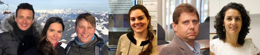
The study, recently published in ACS Applied Materials & Interfaces (Impact Factor 8.097), was developed during the Master’s research work of Wallace Rosado Rolim, guided by Professor Amedea Barozzi Seabra, and defended this year in the postgraduate program in Science and Chemical Technology of the Federal University of ABC (UFABC). The work also involved, through scientific collaborations, knowledge and experimental techniques of Biology and Biomedicine areas. “I emphasize the importance of interdisciplinarity and teamwork for the success of scientific and technological research,” says Professor Seabra, who is the corresponding author of the article.

The idea of developing this biomaterial (a material planned to interact with a biological system for medical diagnostic or treatment) came up in discussions between Rolim and his advisor. “We were looking for new strategies for controlled, localized and sustained release of actives such as nitric oxide molecules associated with silver nanoparticles, for biomedical applications,” reports Professor Seabra. The scientific duo had the idea of bringing together the two therapeutic assets in a single material that was able to topically release them. “We were looking for a synergistic action of these two assets,” says Seabra.

Thus, Professor Seabra and Rolim, with the collaboration of the Master’s student Joana Claudio Pieretti, endeavored to develop the material. The team was able to prepare films made from a composite material, whose matrix consists of a polymer, known as PVA, added with another polymer, called PEG, which made the matrix more flexible. Both polymers are non-toxic and biocompatible. During the preparation of the films, they added silver nanoparticles and a nitric oxide donor substance (the GSNO molecule, which, spontaneously, decompose and generate nitric oxide).

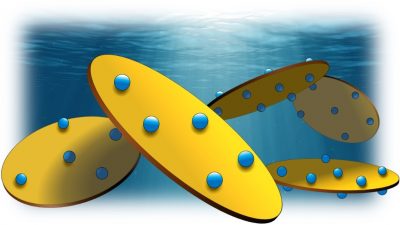
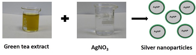
The same group prepared the silver nanoparticles using a simple, inexpensive method that is friendly with the environment and living organisms, also developed by Rolim in his Master’s work. In the method, which was reported in an article published earlier this year (https://doi.org/10.1016/j.apsusc.2018.08.203), green tea extract is used to generate the nanoparticles from silver nitrate, as shown in this figure:


image 1

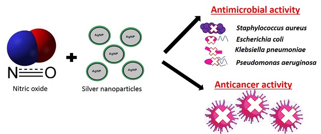
In order to compare the antimicrobial and anticancer effects, the team prepared several types of films: some formed by the pure matrix (PVA/PEG), others containing silver nanoparticles or nitric oxide donors in different concentrations, and the last containing both therapeutic agents in the same matrix. After analyzing all the films using various characterization techniques to accurately determine their composition and morphology, Professor Seabra and her students studied how the release of nitric oxide and silver nanoparticles occurred.

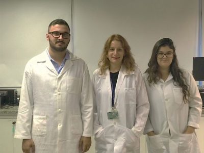
Finally, the films were sent to the collaborators from other research groups to perform the biological assays, which were performed in vitro (i.e., outside living organisms and within environments with controlled conditions). At UFABC, the groups of professors Ana Carolina Santos de Souza Galvão and Christiane Bertachini Lombello focused on the anticancer action of the biomaterial, using cervical and prostate cancer cells. On the other hand, the tests related to the antibacterial activity of the films were carried out at the São Paulo State University of Campinas (UNICAMP), by Professor Marcelo Brocchi’s group, which involved tests with several types of bacteria, including the well-known Escherichia coli and Staphylococus aureus.

The tests showed that the films containing both therapeutic assets presented the best results in the elimination of bacteria and mainly of cancerous cells, as this figure illustrates:


image 2

As a result, the synergism between silver and nitric oxide nanoparticles, which Seabra and Rolim had looked for since the beginning of the Master’s research work, was proven. In one of the assays, to cite one example, less than 25% of the cancer cells remained alive (viable) after being treated with these films for 24 hours.

The material developed by the UFABC team brings the possibility of implementing a new therapeutic strategy for some cancerous tumors and infectious lesions, based on the simultaneous release of nitric oxide and silver nanoparticles, directly at the affected site, from a film. Seabra explains that “In practice, this film can be applied, for example, in a tissue (such as the skin or mucosa) or an organ, for antimicrobial or antitumor actions.” By releasing therapeutic amounts of the agents directly at the site of interest, it avoids unwanted release in healthy organs and/or tissues and thus prevents possible side effects, Seabra adds.

This work received financial support from the Brazilian agencies CNPq, FAPESP and CAPES. The first author of the paper, Wallace Rosado Rolim, developed his master’s research work with a grant from UFABC.

Featured paper: Nanoclays to overcome toxicity.

[Paper: Reaching Biocompatibility with Nanoclays: Eliminating the Cytotoxicity of Ir(III) Complexes. Malte C. Grüner, Kassio P. S. Zanoni, Camila F. Borgognoni, Cristiane C. Melo, Valtencir Zucolotto, and Andrea S. S. de CamargoACS Applied Materials & Interfaces 2018 10 (32), 26830-26834DOI: 10.1021/acsami.8b10842.]

Nanoclays to overcome toxicity

Working in laboratories of the São Carlos Institute of Physics (IFSC – USP), a scientific team developed a strategy that eliminates the cytotoxicity (ability to destroy cells) of a group of compounds with very interesting photophysical properties for health applications . The study made viable the use of these substances, once toxic, in the study of living organisms and in the diagnosis and treatment of diseases. In addition to eliminating cytotoxicity, the strategy modifies some properties of compounds by adding new functions that can be harnessed for intracellular oxygen sensing and to improve the efficiency of luminescent devices such as OLEDs.

The work was reported in an article recently published in the journal ACS Applied Materials and Interfaces (impact factor 8,097).

It all started in an informal conversation between three postdoctoral fellows linked to IFSC-USP laboratories: Malte C. Grüner and Kassio P. S. Zanoni, both linked to the Laboratory of Functional Materials Spectroscopy (LEMAF), and Camila F. Borgognoni of the Group of Nanomedicine and Nanotoxicology (Gnano). Zanoni had worked with iridium (III) complexes during his doctorate, and wanted to take advantage of some properties of these compounds to use them as photodynamic therapy agents. Such therapy refers to a set of treatments for diseased tissues, such as those affected by cancer, in which an external radiation source is used for the activation at the appropriate time of a compound inserted into the body, which is responsible to destroy the cells that need to be eliminated.

The post-doc Zanoni’s desire, however, came up against the high cytotoxicity of iridium (III) complexes. The postdoc Grüner then had the innovative idea of trying to use laponites (materials he had studied in his doctorate) to inhibit the cytotoxicity of the compounds. From this idea, Grüner and Zanoni carried out the preparation and characterization of the materials in LEMAF, coordinated by Prof. Andrea S. S. de Camargo. At GNano, coordinated by Prof. Valtencir Zucolotto, the post-doc Borgognoni and the student Cristiane Melo were in charge to investigate the interactions of the nanoparticles with the cells.

The authors of the paper. From the left: Kassio Zanoni, Camila Borgognoni, Malte Grüner, Cristiane Melo, Valtencir Zucolotto, and Andrea de Camargo.

Strategy and applications

Illustration of the adsorption of Ir (III) complexes (blue spheres) on the surface of laponite nanodisks (yellow disks), in solution.

One of the main properties of iridium (III) complexes is their intense luminescence (emission of light not resulting from heat) in a wide range of colors. This feature may be useful for illuminating cells within living organisms in bioimaging techniques, used for both research and for diagnosis and treatment of diseases.

In turn, laponites, which are synthetic nanoclays fully compatible with living tissues, have often been proposed in the scientific literature as nanoplataforms for transporting drugs and other compounds within living organisms. The laponites are about 25 nm in length and only 1 nm in height.

In the work of the IFSC-USP team, a new material was developed as a result of the adsorption of iridium (III) complex molecules on the surface of laponite nanodiscs.

The researchers found in the laboratory (in vitro) the ability of the new material to be absorbed by cells, its luminescence within cells and its low citotoxicity. For this, they used liver cells and observed their interaction with the new nanomaterial, comparing it with the interaction with the pure iridium (III) complex. The results were highly favorable to iridium (III) laponite nanodiscs, which proved to be harmless to the cells, besides presenting good penetration and high luminescence – characteristics that make them very suitable for application in bioimaging techniques.

Light emission in various colors of the developed nanomaterials (Ir (III) complexes adsorbed on laponite) distributed in xerogels (upper part) and in liver tissue cells (lower part).

“In this work, it was demonstrated for the first time that the adsorption of iridium (III) complexes (in general, highly toxic) on the surface of laponite nanodisks is capable to completely extinguish the cytotoxicity of these compounds “, summarizes the post-doc Kassio Zanoni , who in 2017 was the winner of B-MRS Young Researcher Award. “This makes it highly feasible to use previously toxic compounds in cell media without impairing the integrity of the medium and therefore has the potential to expand the research of new biocompatible materials for use in cell mapping, theranostics and photodynamic therapy”, he adds.

According to the authors, the new nanomaterial could act as a photodynamic therapy drug, since, when irradiated with certain types of radiation, it produces a molecule (the singlet oxygen) that acts in the destruction of cancer cells. In this way, the nanomaterial also becomes promising in the field of theranostics, which proposes the combination, on the same platform, of the diagnosis of diseases by bioimaging with its cure through photodynamic therapies.

In addition, the nanomaterial can be used as a sensor to accurately determine the amount of oxygen distributed inside a cell. “As demonstrated in our work, the emission intensity of this nanomaterial is a variable as a function of the concentration of oxygen”, justifies Zanoni.

Finally, the nanomaterial, in the form of a thin nanometric film, could also be applied to organic light-emitting diodes (OLEDs) – devices that are already used, for example, in cellular screens. “This is because the iridium (III) complex adsorbed on laponite aggregates photophysical, photochemical and electrochemical properties that are strategic for the development of more efficient devices”, explains Zanoni.

This research was carried out with funding from The São Paulo Research Foundation (FAPESP).

People in SBPMat community: SBPMat council member Carlos Roberto Grandini and three other researchers from Brazil integrate the international college of fellows of the International Union of Biomaterials Societies.

Brazilian researchers were awarded the status of “Fellow, Biomaterials Science and Engineering” (FBSE) by the International Union of Biomaterials Science (World’s Biomaterials Societies). The honorary title is a recognition of excellence in professional performance and the achievements made in the field of Biomaterials Science and Engineering. The new fellows now are part of an international college, joining 300 researchers who are committed to strengthening and divulging the field of Biomaterials. The honor was awarded in a ceremony at the opening of the 10th World Biomaterials Congress, held in Montréal (Canada) in May.

One of the new FBSE of Brazil is Professor Carlos Roberto Grandini (from the State University of São Paulo, UNESP, campus Bauru), counselor of SBPMat and 1st Vice President of the Latin American Society of Biomaterials and Artificial Organs (SLABO). Grandini received the honorary title for his contributions in the field of metallic biomaterials and for his leadership in the Latin American scientific community. Besides Grandini, the other new fellows are the Brazilian researchers Aron Jose Pazin de Andrade (Instituto Dante Pazzanese de Cardiologia), Luís Alberto Loureiro dos Santos (Federal University of Rio Grande do Sul, UFRGS) and Marivalda de Magalhaes Pereira (Federal University of Minas Gerais, UFMG).

Professor Grandini receiving the title of “Fellow, Biomaterials Science and Engineering”.

Capes Doctoral Dissertation Award 2015: Interview with the author of the award-winning work in Materials area.

Edroaldo Lummertz da Rocha, winner of the Capes Dissertation Award in Materials field.

The scientific career of Edroaldo Lummertz da Rocha is permeated by two features of the area of Materials science and technology: interdisciplinarity and impact on people’s lives.

After graduating in Computer Science from Universidade do Extremo Sul Catarinense (UNESC), in the south of Brazil, Edroaldo got his master’s degree in Electrical Engineering at the Federal University of Santa Catarina (UFSC), with a study on the transport of phonons in fractal geometry. In 2010, he began a doctoratal program in Materials Science and Engineering, also at UFSC. With the advised of Professor Carlos Renato Rambo and Professor Luismar Marques Porto, Edroaldo worked on the interface among Materials Science, Computer Science and Biology to study the interaction between biological cells and bio and nanomaterials. In 2012, Edroaldo submitted, as first author, a scientific paper on simulations of interactions between nanoparticles and cell membranes. His paper was published in the journal Physical Chemistry Chemical Physics (DOI: 10.1039 / C2CP44035K) in 2013 and was featured on the front cover.

From January to December 2013, Edroaldo remained in the United States developing a part of his doctoral research at Harvard University (United States), more precisely in the Wyss Institute for Biologically Inspired Engineering, a multidisciplinary and multiinstitutional entity dedicated to the development of materials and devices inspired in nature, aimed at transforming medicine and building a more sustainable world. There he was advised by Professor Donald E. Ingber, founder and director of Wyss.

Two papers signed by Edroaldo became covers of prestigious journals.

A second journal cover (DOI:10.1016/j.cell.2014.07 .020) increased Edroaldo´s curriculum in 2014 as a result of his participation, along with researchers at Harvard University and other institutions in the United States, in a Cell Engineering field study.This time the distinction was in Cell, the prestigious journal of Life Sciences, whose impact factor is 32.242.

In September 2014, Edroaldo obtained his PhD degree by defending his dissertation entitled “Nanoparticle-cell interactions and biomaterial-cells induce global changes in gene expression programs“. A year latter, the work was distinguished as the best  dissertation defended in Brazil in 2014 by Capes, the government agency linked to the Brazilian Ministry of Education in charge of promoting high standards for post-graduate courses in Brazil. Edroaldo received the news of the award in the city of Rochester, in the United States, where he works in scientific activities as a postdoctoral fellow of the Mayo Clinic, an institution in the field of Medicine dedicated to research, education and patients´ care.

Interview with Edroaldo.

SBPMat Newsletter: – Could you tell us very briefly how did you become interested in science and in the Materials area, and what were the most important moments in your academic career so far?

Edroaldo Lummertz da Rocha: – My interest in science arose from the possibility of doing something important and helping people somehow. My interest in the Materials area arose due to the existence of a special class of materials, called nanomaterials, which can be used for the development of new therapies for a variety of diseases such as cancer, vascular and neurodegenerative diseases. However, the understanding of how nanomaterials interact with cells and biological tissues is extremely important for the development of safe and effective therapies.

The first most important event of my academic career was when Professor Carlos Renato Rambo, of the Federal University of Santa Catarina, agreed to be my advisor during my doctoral period. That’s where it all began. The second most important moment was when I had the opportunity to conduct part of my doctorate studies at the Wyss Institute at Harvard University, which significantly contributed to the development of my academic career.

Gene regulatory network made of data of gene expression from 16 kinds of human cells and tissues.

SBPMat Newsletter: – What, in your opinion, is the main contribution of your award-winning thesis?

Edroaldo Lummertz da Rocha: – The main contribution of my thesis was the development of computational approaches to systematically understand how cells interact with nanomaterials and respond to external stimuli. This can serve as a basis for future studies in the field of development of new drug delivery systems and lead to a better understanding of how gene expression programs change when nanomaterials interact with cells.

SBPMat Newsletter: – Which criteria guided you to make a quality research highlighted at national level (the award-winning thesis)? To what factors do you attribute this achievement?

Edroaldo Lummertz da Rocha:  – Dedication, discipline, creativity and a good team and co-workers are essential to progress in any area. Family support is essential, above all. From the point of view of the thesis, under the guidance of Professor Carlos Renato Rambo, I had the opportunity to work in a variety of projects and this contributed to the multidisciplinary nature of my thesis.

SBPMat Newsletter: – We invite you to leave a message for our readers who are conducting scientific research in the Materials area.

Edroaldo Lummertz da Rocha: – It is a long road, so you might as well enjoy the way. Being a scientist is something really rewarding, with new challenges and opportunities every day. The hope of discovering something important and providing a significant contribution is the driving force that guides my research. The effort is never in vain and there is always hope where there is perseverance.


More award-winners in the Materials field.

Several other works related to Materials Science and Engineering were awarded this year with the Capes Thesis Award, which was awarded to the best doctoral theses in 2014 in each of the 48 areas of knowledge recognized by Capes in postgraduate courses. The announcement of the winners was made on August 31 and the awards event will take place on December 10 in Brasilia city, the capital of Brazil. Here follow some examples related to Materials area:

Honorable Mention in the Materials area. Thiers Massami Uehara. Study of the interaction of nanomaterials with models of cell membranes and neural stem cells. Advisor: Valtencir Zucolotto. Postgraduate Program in Science and Engineering of Materials – USP/SC. Dissertation file: http://www.teses.usp.br/teses/disponiveis/18/18158/tde-27102014-134646/pt-br.php

Capes Award in Chemistry.  Rodrigo Villegas Salvatierra. Thin Films of Conjugated Polymer and Carbon Nanostructures obtained in Liquid-Liquid Interfaces: Synthesis, characterization and application in photovoltaic devices. Advisor: Aldo José Gorgatti Zarbin. Postgraduate Program in Chemistry – UFPR. Dissertation file:   http://dspace.c3sl.ufpr.br:8080/dspace/handle/1884/37915

Honorable mention in Chemistry.  Anderson dos Reis Albuquerque. Quantum-Chemical Study of the Ti(1-x)CexO2-δ in the Anatase Phase. Advisors: Ieda Maria Garcia dos Santos (DQ-UFPB) and Júlio Ricardo Sambrano (DM-UNESP Bauru). Postgraduate Program in Chemistry – UFPB. Thesis file: http://tede.biblioteca.ufpb.br/handle/tede/7154?locale=pt_BR. Report on CDMF website: http://www.nanotecnologia.com.br/trabalho-orientado-por-professor-do-cdmf-recebe-mencao-honrosa-no-premio-capes-2015/

Featured paper: Promising composite biomaterial for regeneration of bone tissue.

[Paper: Assisted deposition of nano-hydroxyapatite onto exfoliated carbon nanotube oxide scaffolds. Hudson Zanin, Cintia M. R. Rosa, Noam Eliaz, Paul W. May, Fernanda Roberta Marciano and Anderson O. Lobo. Nanoscale, v. 7, p. 10218-10232, 2015. DOI: 10.1039/C4NR07317G]

In a study conducted by the Laboratory of Biomedical Nanotechnology (NANOBIO) from the Brazilian University of Vale do Paraíba (UniVap), in a partnership with scientists from the Brazilian National Institute for Space Research (INPE) and universities from Israel and the UK, a biomaterial was produced that advanced the understanding of some stages on the generation of bone tissue in vitro (out of the real biological context). The low cost material has the potential to be used to accelerate the regeneration of bone tissue in vivo – which may be useful in case of bone fractures, for example. The results of the study were recently published on the scientific journal Nanoscale, from the Royal Society of Chemistry.

In broad terms, the natural generation of bone tissue occurs when cells called osteoblasts produce the organic part of bones to later cover it with the inorganic part, the hydroxyapatite – chemical formula Ca5(PO4)3(OH). The natural deposition of hydroxyapatite is a biomineralization process (production of minerals by living organisms). The biomineralization is not yet fully understood, but its comprehension is of great importance for the development of applications aiming to create bone tissue or firm implants into bones.

“The paper contributes to the understanding of the precipitation process of carbonated hydroxyapatite in vitro, for short amounts of time, over three-dimensional surfaces based on nanohydroxyapatite, vertically aligned carbon nanotubes and graphene”, says Professor Anderson de Oliveira Lobo, Biomedical Engineer with master’s and doctoral degrees in Materials Physics and Chemistry, who signs the paper as its corresponding author.

In association with the Diamonds and Related Materials (DIMARE) group from INPE, represented by researcher Evaldo José Corat, the NANOBIO team, coordinated by Professors Anderson de Oliveira Lobo and Fernanda Roberta Marciano, produced scaffolds with vertically aligned carbon nanotubes. The nanotubes underwent a process of surface oxidation by the plasma etching technique, which exfoliated their tips, produced graphene oxide and thus created a more favorable environment for the formation of nanohydroxyapatite nuclei in the following production stage of the material, the electrodeposition.  This deposition technique was chosen because, among the known methods, it produces the artificial apatite most similar to the biological one, in terms of microstructure and dimensions. In order to set the electrodeposition parameters by means of electrochemical studies, the researchers from Brazil requested the collaboration of a specialist on the subject at a global level, scientist Noam Eliaz, from the University of TelAviv. After the electrodeposition, the team obtained a composite material which retained the biological properties of hydroxyapatite, with the advantage that the presence of the carbon nanotubes reinforced the hardness and resistance of the material.

Afterwards, the scientists submerged the material in simulated body fluid (a liquid that simulates the conditions of blood plasma and is commonly used in biomaterials studies). Under these conditions, the composite material, which is bioactive in this kind of liquid, spontaneously formed a layer of carbonated hydroxyapatite, which, jointly with the nanotubes scaffold and hydroxyapatite films, produced a new composite material.

The researchers were able to observe and study the whole process of biomineralization within periods of up to 7 days, and, in the paper, they propose models to explain several stages.  At that moment, more precisely in the discussion on the chemical modelling of how the biomineralization of the composite occurs, it was important to count with the participation of researcher Paul May, from the University of Bristol, jointly with Hudson Zanin, researcher from the Laboratory of Energy Supply and Distribution from UniVap, who was conducting postdoctoral studies in the British university.

Schematics showing the whole production process of nanobiomaterials and the in vitro bioactivity assay. At the top, from left to right, there is: (i) the production of vertically aligned carbon nanotubes, (ii) exfoliation for the exposition of graphene sheets, (iii) a diagram displaying the nanohydroxyapatite electrodeposition process (iv). On the two middle lines it is demonstrated the whole process of biomineralization in vitro, showing how exchanges between cations and anions occur, up to the formation of the carbonated nanohydroxyapatite layer. On the last line, there are micrographs showing the process of biomineralization at the starting time (right after incubation) and after 7 days (last micrograph).

The research went beyond bringing advances to the understanding of biomineralization in vitro. “The comprehension of this in vitro process may be associated to the process of in vivo regeneration of these materials”, says Professor Anderson Lobo. “In vitro studies with human osteoblastic cells and in vivo assays using animals are being carried out by graduate students and postdoctoral fellows at UniVap’s NANOBIO”, he adds.

The origins of the study is found in Lobo’s doctoral research, carried out at the Brazilian Technological Institute of Aeronautics (ITA) and defended in 2011, in which he managed, for the first time, to synthesize composites with vertically aligned carbon nanorods and nanohydroxyapatite. The research counted or counts with the financial support of Brazilian research funding agencies FAPESP, CNPq, FINEP and CAPES.

Biomaterials in event hold by UC from Bauru.

Researchers from Portugal and USA lectured in the 7 th meeting of the UC from Bauru.

The University Chapter Biomaterials (UCB), headquartered in Bauru (city in the state of Sao Paulo) held its seventh monthly meeting in the morning of November 19th, in the São Paulo State University (UNESP). In that occasion, two foreign researchers who were in Bauru delivered seminars in the meeting.

Professor Ana Maria Pires Pinto from Universidade de Minho (Portugal) lectured about new concepts for biomedical applications of metallic matrix composites. Next, nanomedicine in situ was the topic of the seminar given by professor Tolou Shokuhfar, from Michigan Technological University (U.S.A.).

“The researchers were in contact with our group and we were highly complimented by them”, says Diego Rafael Nespeque Correa, president of UCB and PhD Researcher in the UNESP Post-graduate program in Materials Science and Technology. “We managed to maintain an environment of substantial scientific discussions and promotion of our group”, he adds.

UCB is of the five current units of the SBPMat University Chapters Program.

UCB Facebook: https://www.facebook.com/universitychapterbiomaterial

About SBPMat University Chapters Progran: http://sbpmat.org.br/en/university-chapters/